Live-cell high-resolution Brillouin microscopy of U2OS cells
Brillouin microscopy adds a new dimension to established imaging methods by providing mechanical contrast in live, label-free samples. These examples with U2OS cells illustrate how the technique complements brightfield and fluorescence channels to reveal intracellular detail.
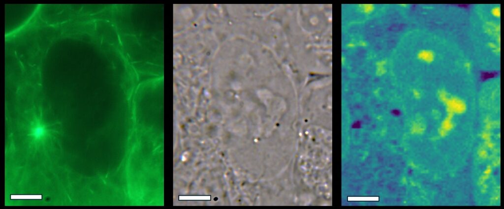
In the first set of images, a single cell is shown in three modalities: fluorescence (left), brightfield (middle), and Brillouin microscopy (right). The Brillouin map highlights differences in intracellular mechanics, with regions of higher water content appearing blue and stiffer regions rendered in yellow. The map is displayed on a frequency shift range of 5.1–5.4 GHz, with a scale bar of 5 µm.
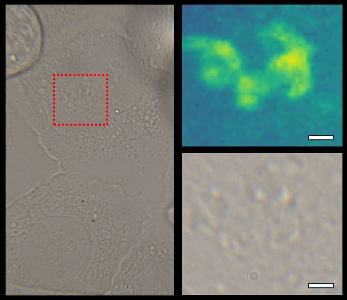
The second set of images presents a brightfield view of U2OS cells with a magnified region (red box) of the nucleus. While conventional techniques provide structural information, the Brillouin map resolves subcellular features that are not readily visible otherwise. A 60× high-NA lens was used, with a scale bar of 2 µm.
Together, these results demonstrate how Brillouin imaging enriches biological insight by adding label-free mechanical information to live-cell studies.
Sample courtesy of Dr. Marketa Schmidt Cernohorska, Centre for Nanomaterials and Biotechnology, Faculty of Science, Jan Evangelista Purkyne University (UJEP), Usti nad Labem, Czech Republic.