Extend Mechanobiology to the volume
From cells to organoids and tissues or small organisms
Mechanobiology is an interdisciplinary field that studies how physical forces and changes in the mechanical properties of cells and tissues influence biology. This area of research delves into how cells sense, respond, and adapt to mechanical stimuli, which can range from fluid flow and pressure changes to the stiffness of the cellular environment. Insights gained from mechanobiology are crucial for understanding disease processes, like cancer progression and cardiovascular diseases, and for advancing regenerative medicine and tissue engineering by manipulating mechanical cues to guide cell behavior and function.
Brillouin Microscopy significantly adds to the mechanobiology toolbox by enabling researchers to non-invasively measure the mechanical properties of cells and tissues in their natural state. This tool can precisely detect stiffness and elasticity at the microscale, providing crucial insights into how cells respond to their mechanical environment. Such capabilities are vital for exploring the physical aspects of cell behavior, which are often involved in critical processes like cell differentiation, migration, and organ development. By facilitating a deeper understanding of these mechanical interactions, Brillouin Microscopy helps uncover the underlying mechanical factors in health and disease, potentially leading to new therapeutic strategies.
Brillouin Microscopy complements modern microscopy techniques by adding valuable new contrast channels to existing imaging systems. Its label-free nature facilitates easy integration with advanced platforms, enabling researchers to correlate mechanical data with chemical and biological insights from other methods such as fluorescence and confocal microscopy. This integration allows for the overlay of mechanical maps on high-resolution cellular images without the need for markers or stains, providing a more complete view of cellular dynamics and interactions across various biological contexts.

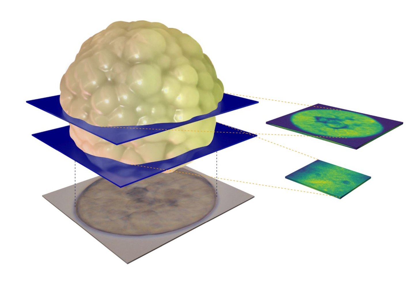
Measuring Mechanics in Regenerating Hydra
The tiny freshwater polyp Hydra vulgaris can regrow an entire body from just a small piece of tissue. To explore how this works, we used Brillouin microscopy for the first time on Hydra and on Hydra spheroids. Because these samples are transparent and alive, the method lets us map their mechanical properties without any labels or preparation.
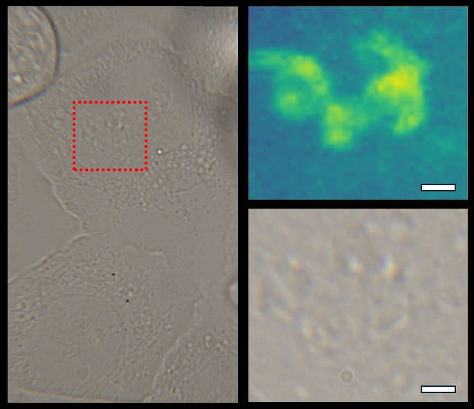
Live-cell high-resolution
Brillouin microscopy adds a new dimension to established imaging methods by providing mechanical contrast in live, label-free samples. These examples with U2OS cells illustrate how the technique complements brightfield and fluorescence channels to reveal intracellular detail.

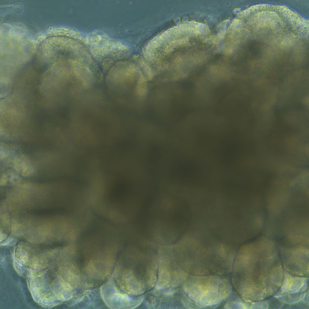
MCF-7 Cancer Organoid Investigations
Brillouin microscopy is revolutionizing cancer research by enabling non-invasive analysis of tumor biomechanics in 3D environments. Unlike traditional methods that require isolating single cells, this technique allows scientists to study live tumor spheroids in physiologically relevant conditions. Recent research demonstrated its potential by investigating MCF-7 breast cancer cells within ECM-mimicking hydrogels. They tracked cell cycle progression while mapping mechanical properties at subcellular resolution. Their findings highlight how tumor cells respond to microenvironmental cues, providing critical insights for precision medicine and the development of targeted cancer therapies.
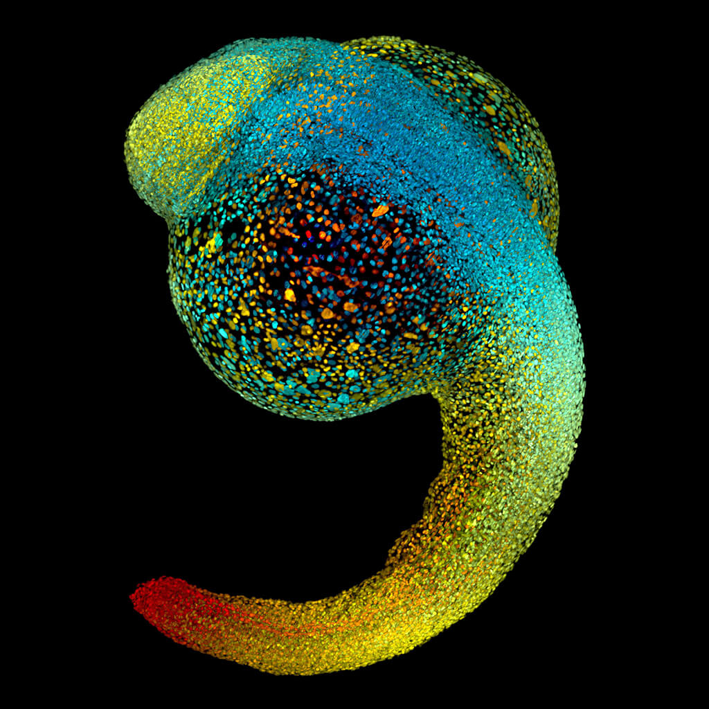
Zebrafish retina
Exploring the mechanical properties of the developing eye in zebrafish larvae in three dimensions. The study uses Brillouin Microscopy to explore the mechanical properties of the developing eye in zebrafish larvae, measuring elasticity and viscosity from eye tissues. It gives insights into the eye’s complex longitudinal modulus, crucial for understanding the mechanobiological aspects of vertebrate eye development.
Cancer organoid
Brillouin Microscopy was used to explore how tumor spheroids’ mechanical properties are influenced by the stiffness and degradability of their 3D microenvironment. Findings reveal that stiffer microenvironments lead to increased spheroid stiffness and reduced invasiveness, highlighting the critical role of mechanical context in cancer progression.
Bladder tissue
Brillouin Microscopy was used to map the mechanical properties of murine bladder tissues, revealing significant differences between healthy and fibrotic conditions. It demonstrates the potential of Brillouin Microscopy as a diagnostic tool to complement traditional histopathological analysis.