Modern biology and medicine
Measure Mechanics in
Uncover mechanical insights in spheroids, organoids and tissues
3D cell culture technology is transforming biomedical research by creating models that closely mimic the natural, in vivo environments of cells. This advanced approach enables the growth of cells in three dimensions, facilitating a deeper understanding of cellular behaviors and interactions compared to the traditional flat, 2D cultures. Utilizing structures such as spheroids and organoids, 3D cell culture allows for the examination of more complex biological processes, including tissue development and organogenesis.
Spheroids, which are aggregates of cells that exhibit near-native tissue organization, are crucial for studying cancer biology and the efficacy of chemotherapeutic agents. Organoids, more sophisticated structures that can mimic entire organs or parts of organs, provide unparalleled insights into developmental biology and disease progression. These 3D models are instrumental in drug testing, disease modeling, and regenerative medicine, significantly enhancing the relevance of in vitro studies.
As 3D cell culture technology continues to advance, it promises to bridge the gap between laboratory research and clinical applications more effectively. The use of these dynamic cell systems is leading to breakthroughs in personalized medicine and targeted therapies, ultimately accelerating the path from laboratory discoveries to clinical solutions. This ongoing evolution not only improves our understanding of human biology but also paves the way for more effective treatments and innovative therapeutic strategies.
Brillouin Microscopy is enhancing 3D cell culture studies by non-invasively measuring the mechanical properties of cells in structures like spheroids and organoids. This method provides insights into the biomechanics of cellular interactions, crucial for replicating human tissue characteristics and improving disease models. With its ability to deliver real-time, label-free imaging, Brillouin Microscopy is a valuable tool in mechanobiology, helping researchers better understand and mimic human tissue behavior in the lab.
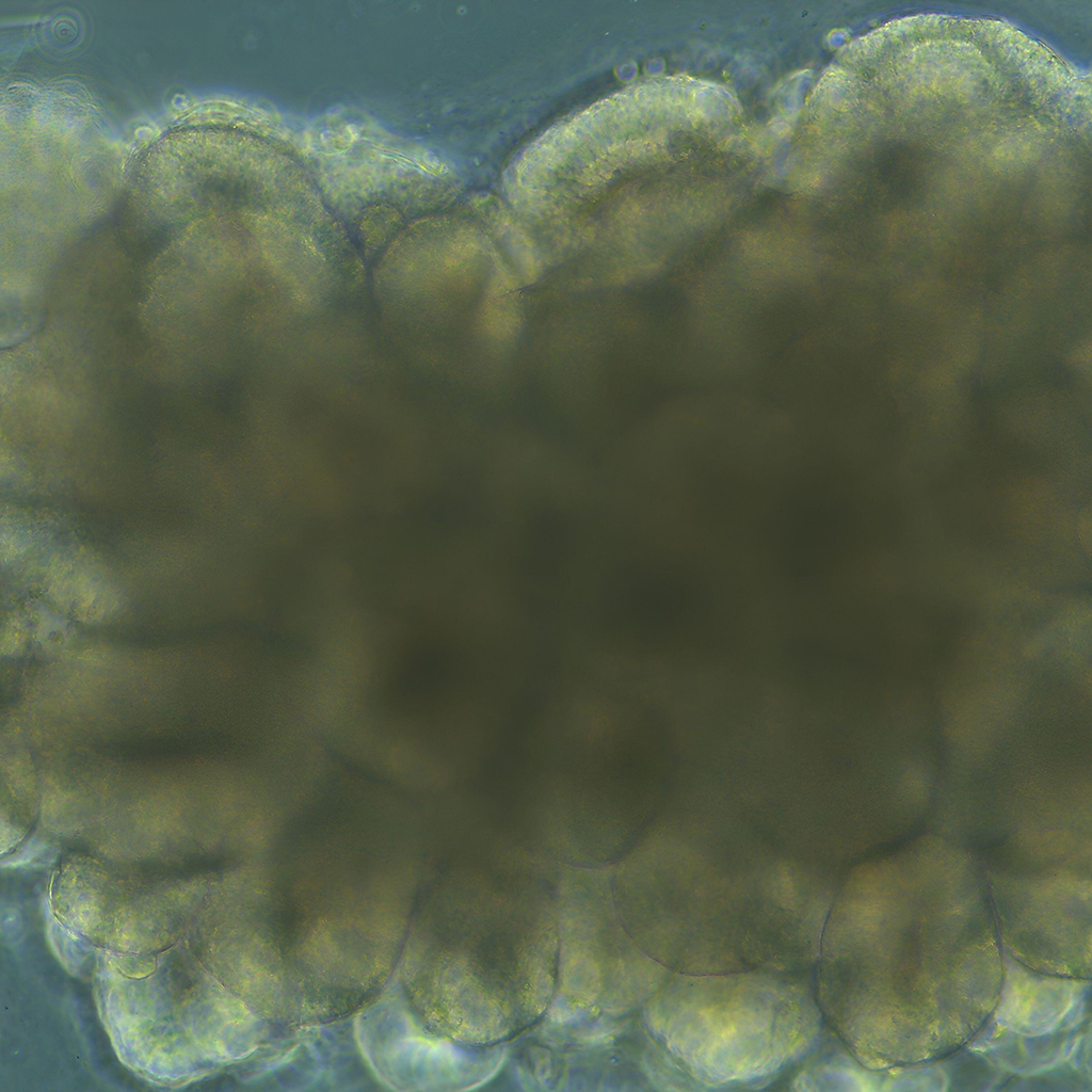
Brillouin microcopy on 3D cellculture models: Cancer organoid
Brillouin Microscopy was used to explore how tumor spheroids’ mechanical properties are influenced by the stiffness and degradability of their 3D microenvironment. Findings reveal that stiffer microenvironments lead to increased spheroid stiffness and reduced invasiveness, highlighting the critical role of mechanical context in cancer progression.