Measuring Mechanics in Regenerating Hydra
The freshwater polyp “Hydra vulgaris” is a classical model for regeneration studies. Despite its simple body plan – a tube with a head and tentacles at one end and a foot at the other—it exhibits remarkable developmental plasticity. Small tissue fragments can regenerate into a complete animal, making Hydra a powerful system for uncovering the interplay between biochemical and mechanical processes in morphogenesis.
Small fragments of Hydra tissue can fold into hollow “spheroids” that can regenerate the complete Hydra body axis. These miniature systems are especially intriguing because they self-organize new body axes and organizers, providing a controlled way to study fundamental principles of tissue patterning and self-organization.
We performed the first Brillouin microscopy measurements on Hydra and Hydra spheroids. Because both are largely transparent and highly hydrated, they are ideally suited for this type of mechanical imaging. Brillouin microscopy directly probes local viscoelastic properties in intact, living samples – an approach that complements molecular readouts and reveals how mechanics guide regeneration. The ability to measure intracellular and tissue-level stiffness without staining or sectioning makes Brillouin an ideal tool for studying such delicate systems.
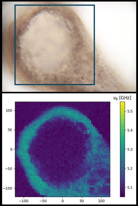
For whole animals, only the region close to the glass surface—the foot of the Hydra—is readily accessible for Brillouin imaging. Measurements in this area reveal the circular symmetry and hollow structure of the hydrozoan. The inner cavity shows the same contrast as the surrounding medium (water), while the cellular layers appear stiffer, with substructures and individual cells becoming visible. A brightfield transmission image marks the measurement region
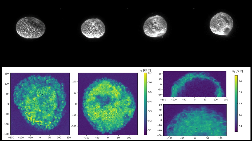
Live spheroids were embedded in a soft hydrogel for immobilization. Brillouin maps of different spheroids revealed striking differences in apparent rigidity and internal organization. A series of fluorescence microscopy images documents Hydra spheroids at various developmental stages, illustrating how mechanical properties evolve during regeneration.
Sample courtesy: Sera Weevers, Friedrich Miescher Institute for Biomedical Research, Basel, Switzerland